
Ophthalmology
Dry eye disease in dogs
![]() Published on March 25, 2025
Published on March 25, 2025
Powered by
![]()
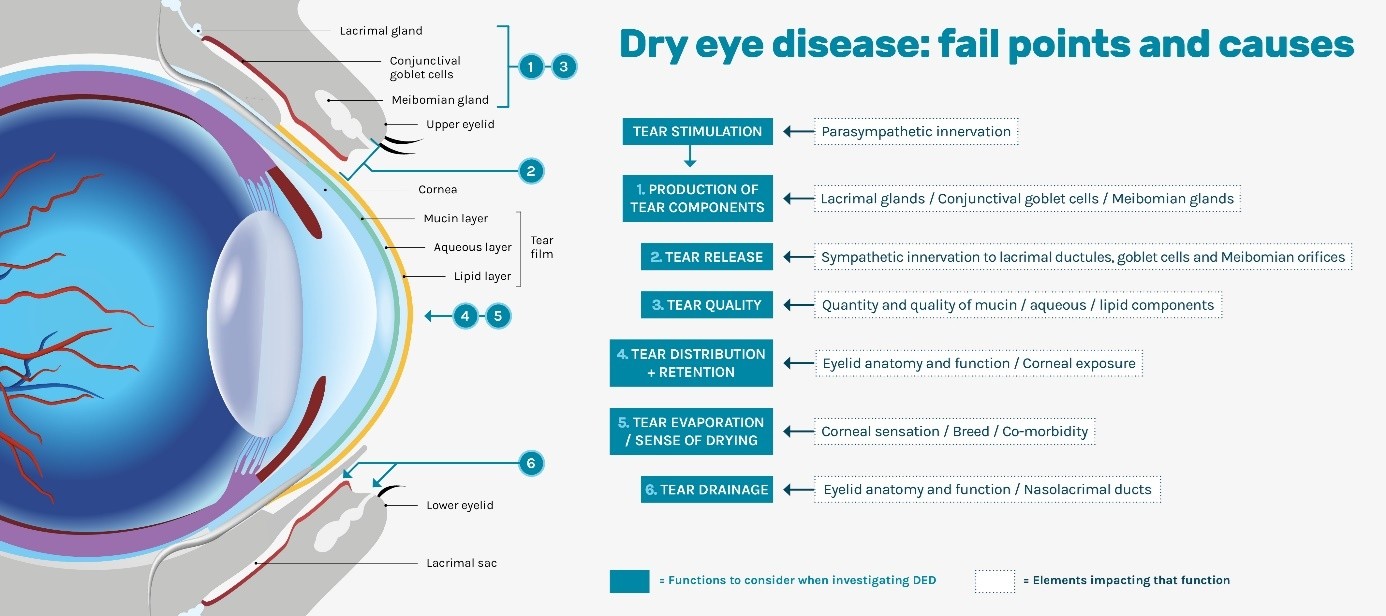
Dry eye disease (DED, keratoconjunctivitis sicca/KCS) is a disease syndrome of the ocular surface arising from dysfunction of the integrated functional system of lacrimal glands, ocular surface, eyelids and/or the motor and sensory nerve supply, which create and distribute the composite preocular tear film. There is accompanying ocular surface inflammation, discomfort, and damage, which can be very mild to very severe.
We tend to place DED into one of three main camps:
- Quantitative (reduced aqueous tear production)
- Qualitative (issues with the mucin or lipid layers of the tear film)
- and/or Distributive (issues with the eyelids).
Modified from (Tear Film and Ocular Surface Society 2007).
The normal tear production and drainage involves a variety of structures and physiological functions. It both requires and preserves the integrity of the ocular surfaces (cornea, conjunctiva).
Single or combined dysfunctions or injuries of either of the structures and functions involved in tear production and drainage can alter the quality or quantity of the tear film and potentially lead to dry eye disease.
The Figure below presents the fail points and causes that can lead to dry eye disease.
Aetiology
Various aetiologies have been described in dogs: developmental, autoimmune adenitis of glandular tissue (the majority of cases of canine DED), metabolic (diabetes, hypothyroidism…), neoplastic, neurologic, infectious, iatrogenic, traumatic or toxic.
Some risk factors have been clearly identified as connected to DED: breed such as brachycephalic dogs (Bulldog, Pug, Lhassa Apso, Shih-Tzu…), age, systemic diseases, medical and surgical history.
|
|
|
|

Treatment & follow-up
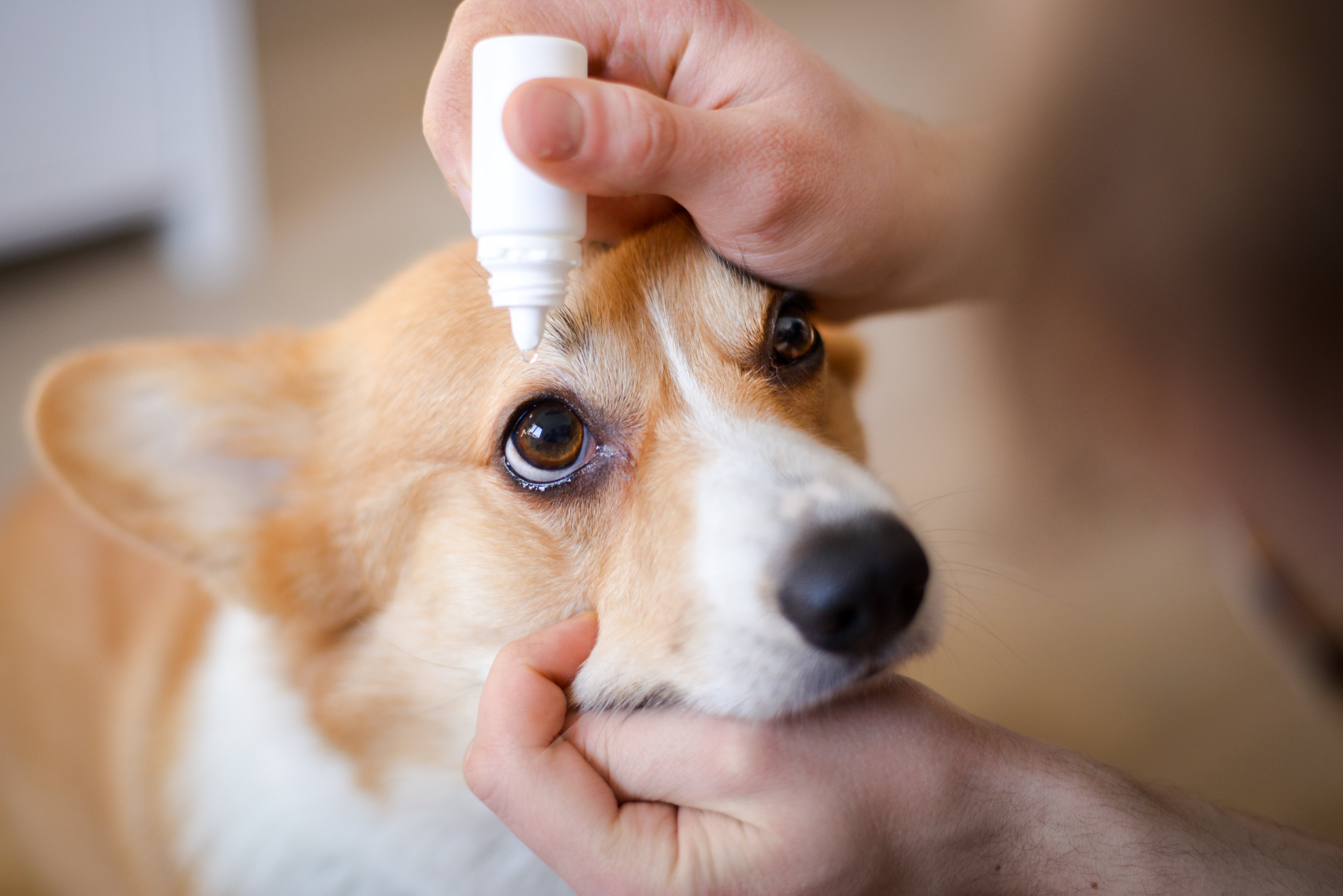
Cyclosporine A and tear supplements (hyaluronic acid…) should always be used as a 1st line treatment.
Topical application of Cyclosporine A every 12 hours is recommended, after cleaning the eye.
Follow-up should be scheduled 1 month after initiation of treatment.
In the absence of response to 1st line treatment, addressing your patient to a referral centre is advised.
Involvement of the owner is essential in the management of DED: teaching them good practices of eye cleaning and treatment administration is essential. Make sure they understand improvement can take several weeks, and treatment can be continued for life.
Eye protection with lubricants and advanced BioHAnce TM technology
Discover an innovation that revolutionizes hydration and eye protection.
You must be logged in to access the page.
The content of this page is based on a guideline on Dry Eye Disease management written collaboratively by:
- Dr Rachael Grundon, Bsc, VetMB, CertVR, CertVOphthal, MANZCVS (Surgery), FANZCVS (Ophthal), DipECVO, MRCVS
- Dr Maria-Christine Fischer, DipECVO, FHEA, MRCVS
- Dr Jens Fritsche, DipECVO
- Dr Alexandre Guyonnet, DipECVO
- Dr Fernando Laguna Sanz, DipECVO